

How Does The Cell Membrane Function In An Animal Cell
With few exceptions, cellular membranes — including plasma membranes and internal membranes — are fabricated of glycerophospholipids, molecules composed of glycerol, a phosphate group, and two fatty acid chains. Glycerol is a three-carbon molecule that functions as the backbone of these membrane lipids. Within an private glycerophospholipid, fatty acids are attached to the first and second carbons, and the phosphate group is attached to the third carbon of the glycerol backbone. Variable head groups are attached to the phosphate. Space-filling models of these molecules reveal their cylindrical shape, a geometry that allows glycerophospholipids to align side-by-side to class broad sheets (Effigy ane).
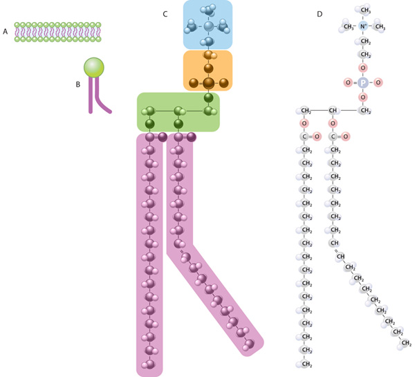
Figure 1: The lipid bilayer and the structure and limerick of a glycerophospholipid molecule
(A) The plasma membrane of a prison cell is a bilayer of glycerophospholipid molecules. (B) A single glycerophospholipid molecule is equanimous of two major regions: a hydrophilic head (green) and hydrophobic tails (royal). (C) The subregions of a glycerophospholipid molecule; phosphatidylcholine is shown as an instance. The hydrophilic head is composed of a choline structure (blue) and a phosphate (orange). This head is connected to a glycerol (green) with ii hydrophobic tails (purple) called fatty acids. (D) This view shows the specific atoms within the various subregions of the phosphatidylcholine molecule. Note that a double bond betwixt 2 of the carbon atoms in one of the hydrocarbon (fat acid) tails causes a slight kink on this molecule, so it appears bent.
Glycerophospholipids are by far the most arable lipids in jail cell membranes. Similar all lipids, they are insoluble in water, simply their unique geometry causes them to aggregate into bilayers without any free energy input. This is because they are two-faced molecules, with hydrophilic (water-loving) phosphate heads and hydrophobic (water-fearing) hydrocarbon tails of fatty acids. In h2o, these molecules spontaneously marshal — with their heads facing outward and their tails lining up in the bilayer's interior. Thus, the hydrophilic heads of the glycerophospholipids in a cell's plasma membrane face up both the water-based cytoplasm and the exterior of the prison cell.
Altogether, lipids account for about half the mass of cell membranes. Cholesterol molecules, although less abundant than glycerophospholipids, account for nigh xx percentage of the lipids in animal cell plasma membranes. However, cholesterol is not present in bacterial membranes or mitochondrial membranes. As well, cholesterol helps regulate the stiffness of membranes, while other less prominent lipids play roles in cell signaling and cell recognition.
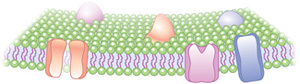
Figure 2: The glycerophospholipid bilayer with embedded transmembrane proteins
In addition to lipids, membranes are loaded with proteins. In fact, proteins account for roughly half the mass of nearly cellular membranes. Many of these proteins are embedded into the membrane and stick out on both sides; these are called transmembrane proteins. The portions of these proteins that are nested amidst the hydrocarbon tails have hydrophobic surface characteristics, and the parts that stick out are hydrophilic (Figure 2).
At physiological temperatures, cell membranes are fluid; at libation temperatures, they get gel-similar. Scientists who model membrane structure and dynamics draw the membrane equally a fluid mosaic in which transmembrane proteins can motion laterally in the lipid bilayer. Therefore, the collection of lipids and proteins that brand up a cellular membrane relies on natural biophysical properties to form and function. In living cells, nonetheless, many proteins are non gratis to move. They are often anchored in place within the membrane by tethers to proteins outside the cell, cytoskeletal elements inside the cell, or both.
Source: https://www.nature.com/scitable/topicpage/cell-membranes-14052567/
Posted by: taylorthenautist.blogspot.com
0 Response to "How Does The Cell Membrane Function In An Animal Cell"
Post a Comment